
Otoplasty: the best surgical solutions for protruding ears
Many people live with a congenital malformation of the ears, and most of them experience this anatomical feature as a psychological handicap. A source of self-consciousness, whether personally, professionally or from early childhood, protruding ears can be a real day-to-day burden. This is why more and more patients turn to various methods to pin back their ears. Whether through otoplasty, an implant or a moulding device fitted in infancy, it is now possible to correct the issue lastingly with a procedure that is usually simple and minor. Many people also wonder whether it is possible to fix protruding ears without surgery: below we review the best solutions, both surgical and non-surgical, and the practical questions patients ask (age, price, recovery, insurance coverage).
Contents
Otoplasty to pin back protruding ears
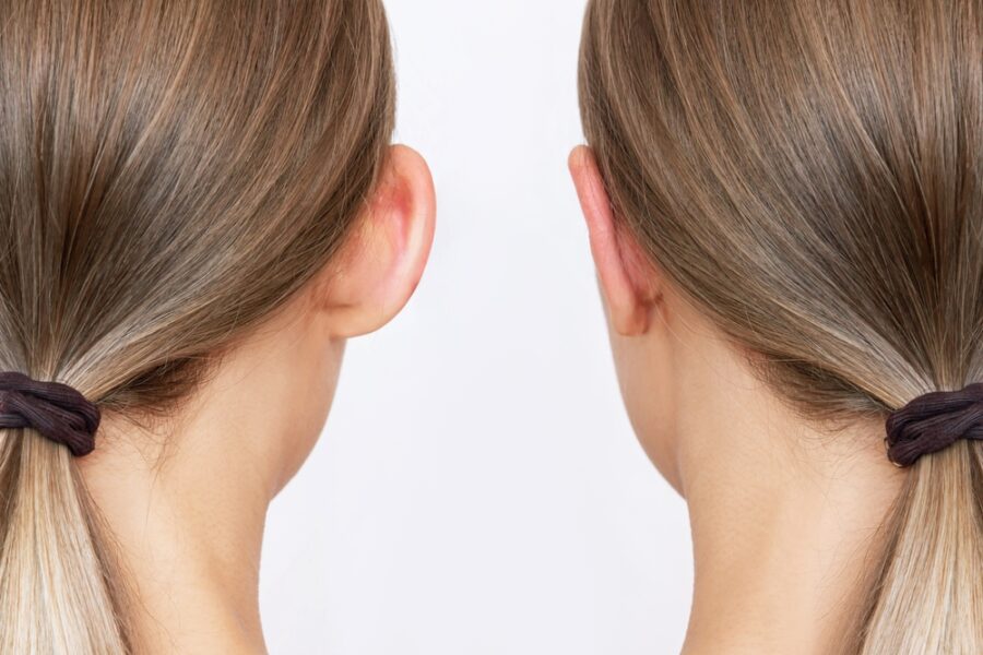
First of all, it should be made clear that protruding ears have no impact on health or hearing. They are, however, an aesthetic concern that can be very hard to live with for the person affected. The teasing that protruding ears can attract may cause psychological distress and a significant drop in self-esteem from a very young age.
The most widely used solution is to pin the ears back with otoplasty. This is a surgical procedure that consists of reshaping the ear cartilage when the ear is protruding. Depending on the nature of the anomalies, the length of the procedure or the patient’s age, the operation can be performed under local anaesthesia, under general anaesthesia or under intravenous sedation.
Indications for otoplasty
Otoplasty, which can be unilateral (one ear) or bilateral (both ears), is a genuine challenge for the surgeon because of the complex surgical and anthropometric anatomy of the ear. Protruding ears are in fact a deformity caused by several anatomical features. These may include:
- A lack of cartilage folding (antihelix), creating an ear that is too smooth and straight and therefore tends to point outwards;
- An overdeveloped cartilage (concha) causing the ear to project too far forwards;
- An abnormal retro-auricular angle creating a gap between the ear and the skull and accentuating the sense of protrusion;
- A valgus of the earlobe, that is, an accentuated protrusion that again gives a distinctive outward-turned shape.
How the procedure unfolds
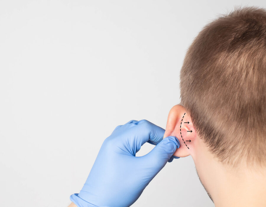
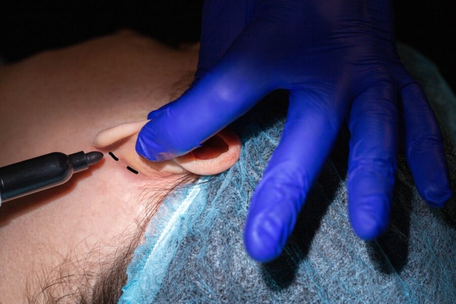
First, the surgeon carries out an examination of the ears as well as a photographic assessment. A pre-anaesthesia work-up is also performed. During the operation, the surgeon makes a skin incision in the retroauricular fold, which corresponds to the crease behind the ear. Slight incisions may also be needed on the front of the ear. The surgeon then lifts the skin over a larger or smaller area, as required, to access the cartilage.
Depending on the issue identified, the surgeon will then use one of the following techniques, some of which are sometimes combined when the malformation requires it:
- Cartilage reshaping by chondrotomy (removal) to redraw the contour;
- Folding of the upper part of the ear (antihelix) to tuck in the concha;
- Correction of the shape of the lobes;
- Removal of a strip of excess skin to avoid creating an unsightly bulge.
After the operation, surgery for protruding ears produces ears that are pinned back from the day after the procedure, with a very natural appearance. The results are even more visible once the swelling subsides after 2 to 3 weeks, with an optimal final result around 3 months.
At what age and under what conditions can ears be pinned back?
Beyond the choice of technique, patients mainly ask practical questions: from what age to operate, what recovery is like, whether the operation is painful and whether it is covered by insurance. Here are the essentials.
The ideal age to pin back the ears
In children, surgeons generally wait until the ear cartilage has completed most of its growth, which happens early: the ear reaches nearly 85 to 90% of its adult size by around the age of 6. This is why otoplasty is most often considered from the age of 7, an age when the ear is almost fully formed and when the child, often confronted with teasing when starting school, asks for it themselves. It remains, of course, possible in adolescence and adulthood, with no upper age limit. In the newborn, conversely, the great malleability of the cartilage allows a non-surgical correction with a moulding device (the Earwell method), provided it is carried out in the very first weeks of life.
Recovery, convalescence and pain
Otoplasty is known for its simple recovery. Most often performed on an outpatient basis, it is not painful during the procedure (under local or general anaesthesia); in the following days, some discomfort and sensitivity are usual and well relieved by standard painkillers. A compression headband is worn continuously for a few days, then at night for several weeks to protect the ears while they heal. Returning to school or work generally happens within a week, with contact sports to be avoided while the cartilage consolidates. The scar, hidden in the crease behind the ear, quickly becomes invisible; as with any incision, careful follow-up helps prevent a thick scar or a keloid scar in skin types that are prone to it.
Price and insurance coverage for otoplasty
When the protrusion is significant and constitutes a genuine burden, otoplasty may qualify for partial insurance coverage by the French health service (Assurance Maladie) after prior approval, particularly in children; the out-of-pocket cost and any additional fees vary depending on the practitioner and the facility. The exact price, which depends on the technique chosen and on whether the procedure is unilateral or bilateral, is given during the consultation, following a detailed quote provided before any decision is made. This same consultation is an opportunity to discuss other procedures to rejuvenate and harmonise the face, such as a facelift, if the patient wishes.
The Earfold technique
Another, even less invasive technique was also available until recently to pin back the ears: the Earfold implant technique. This innovative medical procedure for the permanent correction of protruding ears consisted of inserting a curved metal implant a few millimetres long in front of the ear, at the level of the antihelix fold. Earfold thus offered shape-memory implants made of Nitinol, an alloy of nickel and titanium. This device made it possible to permanently correct the shape of the ears and, above all, the deformity of protruding ears.
The advantages of Earfold
Both simple and quick, this technique was very minimally invasive and represented an effective alternative to conventional surgical methods for patients most reluctant to go under the scalpel. Earfold implants also offered many advantages over otoplasty. The procedure lasted about 20 minutes and took place directly in the practitioner’s office. It was thus performed under local anaesthesia, like dental care. Moreover, it caused no particular recovery apart from slight swelling, and offered immediate initial results, even though the final result was only really visible after a month.
The treated part of the ear had to be left untouched during this consolidation phase, otherwise the implant could shift. Ultimately, this solution could be regarded as otoplasty without surgery in an operating theatre. Children and adolescents with protruding ears could thus benefit from this far less invasive solution than otoplasty.

The limitations of the Earfold technique
It should be noted, however, that this ear-correction technique did not work for every type of patient (around 70% of patients were eligible). It was not indicated when the protrusion involved the concha or when ear cartilage had to be removed. In addition, despite its great discretion, the implant could be slightly visible in patients with relatively thin skin over the ears.
The material used in this technique is unfortunately no longer marketed at present, but the other techniques to pin back the ears nevertheless remain available and very effective.
The Stenström technique
The principle of this technique is to weaken the front surface of the cartilage in a harmonious way. This is done using a curved rasp that follows the curve of the ear. When the cartilage is folded, the weakened area can then take the curve more easily, allowing gentle, harmonious folding of the cartilage. In other words, this method breaks the anterior fibroelastic arch so that the posterior arch provides a centripetal traction force causing the cartilage to roll up spontaneously. This is referred to as tubulisation by partial anterior chondrotomy, that is, a spontaneous rolling-up of the cartilage following its surgical incision.
After identifying the future antihelix, the skin is removed along the axis of the antihelix without lifting the posterior skin. The outer surface of the ear is exposed via an anterior approach limited to the junction between the helix and the upper branch of the antihelix, thus allowing a broad anterior dissection including the antihelix, its branches and the outer part of the concha.
This precision technique requires real experience on the part of the operating surgeon. Different operative methods are used depending on the practitioner. Some perform the anterior dissection at the antitragohelicine fissure (between the tail of the helix and the antitragus). Others fix the tubulisation obtained by partial anterior chondrotomy using sutures, as in the Mustardé technique. Still others perform tucking of the concha and fixing it to the premastoid plane (at the level of the lower part of the temporal bone).
The Earwell method
Specifically dedicated to infants, this neonatal solution aims to act on malformations or deformities of the outer ear present at birth through the placement of a moulding external conformer to correct the shape anomalies of the ear. Ear malformations are in fact very common in babies and are thought to affect 15 to 20% of births, most of them persisting into adulthood with the harmful psychological consequences already discussed above.
During the first 6 weeks of a baby’s life, the cartilage of its ear remains malleable, making correction easier than in adulthood. The ear can therefore be folded more easily when a defect in the ear fold is detected early. Thanks to the Earwell method, it is possible to reposition the newborn’s ear by recreating the missing antihelix fold. This technique is mainly widespread in the United States and is currently practised by some hospitals in France.
Earwell is a moulding technique lasting 4 to 6 weeks, tailored case by case to the type of anomaly. No anaesthesia is required because the conformer is fitted during a simple ENT consultation. This device, which corrects ear deformities in babies atraumatically, therefore requires no surgery and offers a success rate of over 90%. It is thus a non-invasive, non-surgical technique that leaves no scar. The Earwell fitting procedure also takes only a few minutes. Within 4 to 6 weeks, the baby will have ears with a normal shape.
Frequently asked questions
At what age can ears be pinned back?+
Otoplasty is generally offered from the age of 7, once the ear has reached most of its adult size. It remains possible at any age afterwards, in adolescence as in adulthood, with no upper limit. In the newborn, a non-surgical correction with a moulding device (the Earwell method) can be considered in the first few weeks of life, while the cartilage is still very malleable.
Can ears be pinned back without surgery?+
Yes, but it depends on age. In the infant, the Earwell external conformer can reshape a protruding ear without any procedure, while the cartilage is still soft. In adults, by contrast, the cartilage is rigid: no trick, bandage or « home remedy » can lastingly pin back ears that are already formed. The only truly permanent correction is then otoplasty.
Is pinning back the ears painful?+
The procedure itself is not painful, as it is performed under local or general anaesthesia. In the days that follow, some sensitivity and discomfort are usual; they are well relieved by simple painkillers. Wearing a compression headband protects the ears while they heal.
How long is recovery after otoplasty?+
Recovery is straightforward. Returning to school or work generally happens within a week. The headband is worn continuously for a few days, then at night for several weeks. Swelling subsides within 2 to 3 weeks, and the final result can be assessed at around 3 months. Contact sports should be avoided while the cartilage consolidates.
Is otoplasty covered by French health insurance?+
When the protrusion is marked and constitutes a genuine burden, otoplasty may qualify for partial insurance coverage by the French health service (Assurance Maladie), after prior approval, particularly in children. The out-of-pocket cost and any additional fees depend on the practitioner and the facility; a detailed quote is provided during the consultation.
Can the ears come unstuck again after the operation?+
The result of otoplasty is considered lasting. Once the cartilage has been reshaped and stabilised, the ears keep their new position. A slight partial recurrence remains possible in rare cases, most often linked to the quality of the cartilage or to early trauma; wearing the compression headband while the ears heal limits this risk.
Book an appointment
If you are considering a procedure, book an appointment in just a few clicks: our surgeons will answer all your questions and guide you toward a tailored, personalised solution.
Book on Doctolib