01What is dermoscopy?
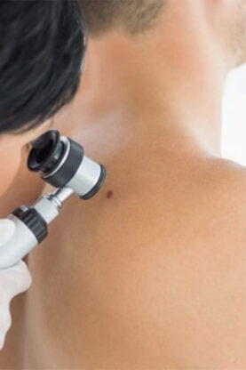
Dermoscopy is a skin examination using a dermatoscope, an instrument fitted with a magnifying lens. Thanks to polarised light (transillumination), it reveals the structure and the vessels inside lesions, which are invisible to the naked eye. The dermatologist examines the entire skin and scalp — a true full-body examination. Every suspicious lesion is photographed and analysed (characteristics, pigmentation, vascular structure); if there is any doubt, it is sent for biopsy.
Because each lesion is catalogued, dermoscopy also makes it possible to map your moles and to monitor each one individually from one session to the next. The examination is non-invasive: perfectly painless, quick and free of side effects.
Its value is proven: in trained hands, the dermatoscope significantly improves the reliability of melanoma diagnosis compared with examination by the naked eye, and avoids the unnecessary removal of benign lesions. It complements two simple guides: the ABCDE rule and the « ugly duckling » sign (the mole that stands out from the others).
02Why have a dermoscopy?
Dermatologists recommend this examination on a regular basis: it is a simple and highly effective way to prevent skin cancer, and an excellent tool for personalised follow-up. It is generally advised every 6 months to 2 years, depending on the number and nature of your moles.
Dermoscopy is also indicated to rule out concern over a specific lesion considered worrying by the patient or their doctor. Finally, it helps with the diagnosis of inflammatory or infectious conditions of the skin, the scalp (scabies, for example), the mucous membranes and the nails. When a lesion turns out to be suspicious, it is removed and then analysed: see mole removal.
03Which skin cancers are detected?
Dermoscopy makes it possible to detect early most malignant skin tumours:
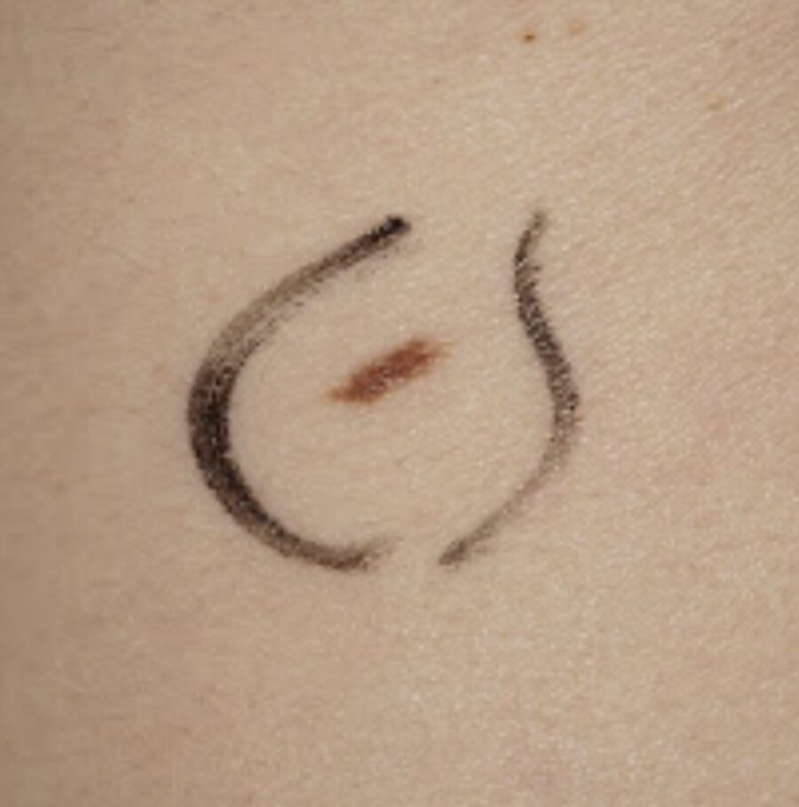
- Melanoma — an aggressive cancer of the melanocytes; the dermatoscope identifies it through asymmetric patterns, varied colours and atypical structures.
- Basal cell carcinoma — the most common skin cancer, generally not very aggressive (see basal cell carcinoma surgery).
- Squamous cell carcinoma — arising from the keratinocytes, sometimes more aggressive than basal cell carcinoma.
- Actinic keratosis — a precancerous lesion of sun-exposed areas that can progress to squamous cell carcinoma.
- Rarer lesions — adnexal tumours, Kaposi’s sarcoma, cutaneous lymphomas, skin metastases.
It also helps to distinguish benign lesions that may change over time (atypical nevi) and to make the differential diagnosis from non-cancerous conditions.
04How the procedure works
Before the procedure
No major preparation is needed. There is just one useful precaution: come without make-up or nail polish, which can interfere with the examination of certain areas. Remember to tell the dermatologist about your medical history (personal or family history of melanoma, sunburn, fair skin type) and to point out the lesions that worry you.
During the procedure
The examination is carried out at the practice, in a single session. The dermatologist runs the digital dermatoscope over the entire skin and scalp (the whole body). Each suspicious lesion is magnified, photographed and recorded — this digital storage allows for mapping and comparison from one session to the next. If there is any doubt, the lesion is sampled for analysis (biopsy). The examination is painless and emits no radiation.
After the procedure
There are no particular precautions after the examination: you resume your activities immediately. The dermatologist gives you their conclusions at the end of the session and, if a lesion has been sampled, the analysis results follow within a few days to two weeks. Depending on your risk profile, a monitoring schedule (6 months to 2 years) is set, with comparative follow-up of the images.
05Prices & fees in Paris
| Procedure | With insurance coverage | Aesthetic fees |
|---|---|---|
| Digital dermoscopy | 120 € | 120 € |
Indicative “from” prices, surgeon fees included. The final quote is given at the consultation, after examination, depending on the area treated and the technique chosen. Part of the procedure may be covered by French national health insurance when the medical criteria are met.
06Your questions
Does dermoscopy examine the whole body?+
Yes. The dermatologist runs the dermatoscope over the entire skin and scalp during the same session. Only the suspicious lesions are then magnified, photographed and analysed in detail.
Is dermoscopy painful or risky?+
No. The examination is completely painless and non-invasive: no aggressive contact, no ionising radiation, no side effects. It can be repeated as often as needed.
How much does dermoscopy cost in Paris?+
At the Rive Droite Paris Étoile practice, the examination costs €120 (see the pricing table below). Insurance coverage is possible for high-risk patients (see below).
Is dermoscopy reimbursed by the French health service?+
It may be covered if you belong to the population at high risk of skin cancer, that is, if: you have already had a melanoma, a first-degree relative has had one, or you have more than 50 moles on your body.
How often should you have a dermoscopy?+
Generally every 6 months to 2 years, depending on the number and nature of your moles and your risk profile. Digital mapping makes it possible to compare images from one session to the next in order to spot any change.
Which dermatoscope is used at the practice?+
A digital dermatoscope, the latest generation: it magnifies and enhances the images with great precision and allows for digital storage of each lesion, for higher-quality detection and comparative follow-up.
What happens if a lesion is suspicious?+
The lesion is photographed and, if there is any doubt, sampled for analysis (biopsy). If it has to be removed, a complete excision is performed, followed by analysis under the microscope: this is the guarantee of a reliable diagnosis.
Is any preparation needed before the examination?+
Very little: come without make-up or nail polish (they interfere with the examination of certain areas) and report your medical history as well as the moles that worry you.